Shrinkwrap - DeformableSurface program
We have developed and implemented a deformable surface model (Computer
Vision and Graphics in Fluorescence Microscopy, LM Lifshitz, KE Fogarty,
JM Gauch, E Moore, SPIE vol. 1808, Visualization in Biomedical Computing,
pp. 521-534, 1992) which has been used numerous times over the years
to locate the plasma membrane of fluorescently labeled smooth muscle
cells. This model is composed of vertices spaced along a model surface
which is initially placed within a 3D image, near its correct location.
Each vertex is then allowed move in response to image intensity gradients,
attempting to move to locations in the image with a higher intensity
(which are typically labeled channels, but possibly other structures such
as locations with strong edge strength). The position of each vertex
also imposes a "cost" based upon how much curvature its position would
impose onto the surface; Vertices are moved to maximize a function of
the image intensity at their location minus a measure of this curvature.
Our implementation calculates curvature measures (i.e., first and second
derivatives of the surface) and intensity measures locally and quickly.
This results in a fast algorithm which is also easy to modify to handle
additional local constraints. For instance, it is simple to freeze
certain vertices in space so they cannot move (e.g., if their initial
position is known to be very accurate) or to restrict motion of the
vertices to a plane (e.g., if a 2D time series is analyzed as one
"3D" image).
Once a surface
(e.g., a plasma membrane 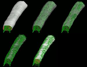 ) has been defined we have numerous
software modules to facilitate analysis of fluorescence relative to
that surface. One module eliminates all signal in an image which is
not within a defined distance from the surface. This has been used
to remove non-specific label inside a cell so that analysis can be
restricted to label which is on, or very near, the plasma membrane.
Another module can map an interior point to the nearest location on
the surface. This enables, for example, finding the distance between a
molecule inside the nucleus and the nuclear membrane (e.g., if it might
be exiting via nuclear pores). Another module creates a histogram of
fluorescent intensity inside a cell as a function of distance from the
plasma membrane (e.g., to enable the application of a threshold which
varies within the cell based upon distance from the plasma membrane).
) has been defined we have numerous
software modules to facilitate analysis of fluorescence relative to
that surface. One module eliminates all signal in an image which is
not within a defined distance from the surface. This has been used
to remove non-specific label inside a cell so that analysis can be
restricted to label which is on, or very near, the plasma membrane.
Another module can map an interior point to the nearest location on
the surface. This enables, for example, finding the distance between a
molecule inside the nucleus and the nuclear membrane (e.g., if it might
be exiting via nuclear pores). Another module creates a histogram of
fluorescent intensity inside a cell as a function of distance from the
plasma membrane (e.g., to enable the application of a threshold which
varies within the cell based upon distance from the plasma membrane).