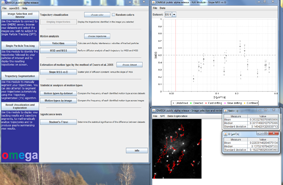
Micro-Meta App is designed to aid in the collection of both Microscope Hardware Specifications and Image Acquisition Settings metadata. In this example, a previously saved Microscope file was selected from an available repository and opened for further editing. In order to add the metadata associated with a newly purchased objective to a Microscope file the “Magnification” drop-down menu is opened [1] and an additional “Objective” [2] is dragged onto the workspace.

Micro-Meta App facilitates the collection of both Microscope Hardware Specifications and Image Acquisition Settings metadata on the basis of the proposed 4DN extension of the OME Core model, which was recently posted as a white paper on ArXiv.org. A beta version of Micro-Meta App was released in December 2019.
Available implementations of Micro-Meta App include:

Background:
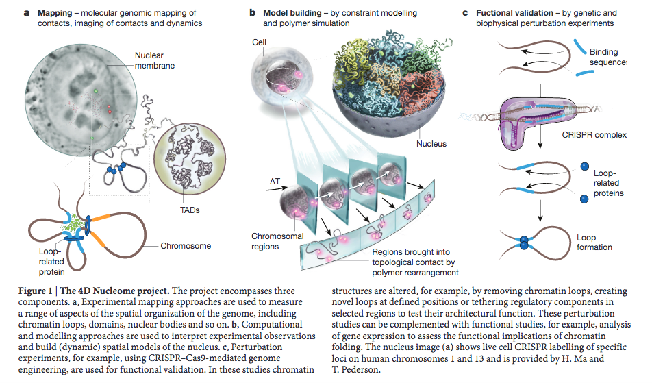
For the information content of image data to be unequivocally extracted and machine-readable, microscopy images need to be accompanied by thorough documentation of the microscope hardware, imaging settings, and instrument performance that ensure the correct interpretation of results. A significant challenge with the reproducibility of microscopy results and with their integration with chromatin folding maps generated by the 4DN Consortium lies in the lack of universally accepted super-resolution microscopy quality control and reporting standards and of widely available cyberinfrastructure to support the collection of provenance metadata. To address this challenge, the 4DN consortium has put forth a 4DN extension of the OME Core metadata model, which includes a tiered system of reporting guidelines that scales quality control and reporting requirements with experimental complexity.

Summary:
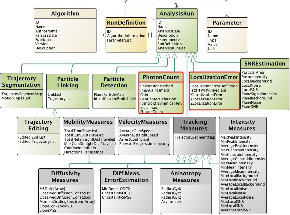
Micro-Meta App is a novel application that provides an interactive and intuitive approach for rigorous record-keeping in fluorescence microscopy and is based on the 4DN-OME Microscopy metadata standard and on the proposed tiered-system of guidelines.  The user’s data processing workflow consists of multiple steps. First, in the Create Microscope modality (Figure 1), the App allows the users to build a graphical representation of the microscope hardware by dragging-and-dropping individual components onto the workspace and entering the relevant attribute values based on the selected tier level. Second, Micro-Meta App generates tier-specific descriptions of the microscope hardware and exports them in a Microscope file that can be used as a template and shared with the community, with a significant reduction in the recordkeeping burden. Then, in the Use Microscope modality, Micro-Meta App opens an existing Microscope file, imports Image Acquisition settings from the header of image data files to be annotated and interactively guides the user through the collection of all missing instrument-specific and tier-appropriate image acquisition settings and calibration metrics required to ensure reuse and reproducibility of image data. Finally, the App generates JSON files that contain comprehensive descriptions of the conditions utilized to produce individual microscopy datasets, and that can be stored on the user’s file system, or on third party repositories. To lower the barrier of adoption of Micro-Meta App by a wide community of users the application is available as a stand-alone program, as a plugin of the OMERO web client and as a service of the 4DN data portal.

In October 2018, Caterina became an associate member of the 4D Nucleome initiative with a project aimed at developing microscopy metadata and particle tracking standards to aide in the reproducibility of shared 4DN imaging datasets.

For more details about the aims of the 4DN project take a look here –>

From: Dekker et al. The 4D Nucleome project. Nature, 549: 219–226 (2017)


A manuscript describing the method we developed to quantify motion type estimation uncertainty was recently published on BiorXiv.org. For more details see here:

Summary:

Quantitative analysis of microscopy images is ideally suited for understanding the functional biological correlates of individual molecular species identified by one of the several available ‘omics’ techniques. Due to advances in fluorescent labeling, microscopy engineering, and image processing, it is now possible to routinely observe and quantitatively analyze at the high temporal and spatial resolution the real-time behavior of thousands of individual cellular structures as they perform their functional task inside living systems. Despite the central role of microscopic imaging in modern biology, unbiased inference, valid interpretation, scientific reproducibility and results dissemination are hampered by the still prevalent need for subjective interpretation of image data and by the limited attention given to the quantitative assessment and reporting of the error associated with each measurement or calculation, and on its effect on downstream analysis steps (i.e., error propagation). One of the mainstays of bioimage analysis is represented by single-particle tracking (SPT), which coupled with the mathematical analysis of trajectories and with the interpretative modeling of motion modalities, is of key importance for the quantitative understanding of the heterogeneous intracellular dynamic behavior of fluorescently labeled individual cellular structures, vesicles, viral particles and single-molecules. Despite substantial advances, the evaluation of analytical error propagation through SPT and motion analysis pipelines is absent from most available tools (Sbalzarini, 2016). This severely hinders the critical evaluation, comparison, reproducibility and integration of results emerging from different laboratories, at different times, under different experimental conditions and using different model systems. Here we describe a novel, algorithmic-centric, Monte Carlo method to assess the effect of experimental parameters such as signal to noise ratio (SNR), particle detection error, trajectory length, and the diffusivity characteristics of the moving particle on the uncertainty associated with motion type classification. The method is easily extensible to a wide variety of SPT algorithms, is made widely available via its implementation in our Open Microscopy Environment inteGrated Analysis (OMEGA) software tool for the management and analysis of tracking data, and forms an integral part of our Minimum Information About Particle Tracking Experiments (MIAPTE) data model.

A manuscript describing the OMEGA application was recently published on BiorXiv.org. For more details see:

Summary:

Open Microscopy Environment inteGrated Analysis (OMEGA) is a cross-platform data management, analysis, and visualization system, for particle tracking data, with particular emphasis on results from viral and vesicular trafficking experiments. OMEGA provides intuitive graphical interfaces to implement integrated particle tracking and motion analysis workflows while providing easy to use facilities to automatically keep track of error propagation, harvest data provenance and ensure the persistence of analysis results and metadata. Specifically, OMEGA: 1) imports image data and metadata from data management tools such as the Open Microscopy Environment Remote Objects (OMERO; Allan et al., 2012); 2) tracks intracellular particles movement; 3) facilitates parameter optimization and trajectory results inspection and validation; 4) performs downstream trajectory analysis and motion type classification; 5) estimates the uncertainty propagating through the motion analysis pipeline; and, 6) facilitates storage and dissemination of analysis results, and analysis definition metadata, on the basis of our newly proposed FAIRsharing.org complainant Minimum Information About Particle Tracking Experiments (MIAPTE; (Rigano and Strambio-De-Castillia, 2016; 2017) guidelines in combination with the OME-XML data model (Goldberg et al., 2005). In so doing, OMEGA maintains a persistent link between raw image data, intermediate analysis steps, the overall analysis output, and all necessary metadata to repeat the analysis process and reproduce its results.

Our proposal for a Minimum Information About Particle Tracking Experiments standard was published on BiorXiv.org. For more details see: biorxiv 155036

Summary:

MIAPTE aims at standardizing the process used to extract particle trajectory data from time series and to analyze their motion as well as providing community guidelines for reporting trajectory data and motion analysis results. In order to serve the needs of a diverse set of possible users, MIAPTE guidelines are provided in three formats:
1) A glossary in tabular format, where data elements are presented together with their properties, their definitions, their cardinality, requirement level, as well as recommended sources for their annotation, examples, and notes (Glossary of MIAPTE guidelines).
2) ER diagrams, which depict each entity together with its attributes as well as the relationship between one another (Section 1 and Section 2).
3) XML schema file, which was created based on our implementation of the MIAPTE guidelines in OMEGA. Such schema can be used to generate the corresponding data structures (Section 1 and Section 2).

Please leave a comment below to help us extend and improve the model.

MIAPTE analysis elements: Entity-Relationship diagram displaying the elements of MIAPTE describing trajectory analysis procedures and results.

In June 2016 we released on fairsharing.org a proposal for a novel minimum information reporting guidelines for Multiple Particle Tracking experiments called  Minimum Information About Particle Tracking Experiments (MIAPTE).

The MIAPTE metadata definition standard is fully OME-XML compatible metadata and we hope it will facilitate the sharing and dissemination of results obtained from Multiple Particle Tracking (MPT) experiments. Specifically, we hope that MIAPTE will contribute to make data produced in MPT experiments FAIR (Findable Accessible Interoperable and Reusable). More info can be found here.

In order to refine and extend MIAPTE we are soliciting comments, suggestions, criticisms from the community.

Alex presented a lighting talk in the morning of the first day. Alex also presented a poster in the afternoon entitled “OMEGA: an open source environment to facilitate the execution of motion analysis workflows, to estimate error and to share result”.

OMEGA motion type classification module

OMEGA motion type classification module

Thanks to the work led by Caterina and Ivo, financially supported by Eric and conducted by Vanni, Alex and Tiziano, a method to determine the motion type of trajectory segments and to automatically compute the prediction error associated with motion type classification, has been integrated into OMEGA. Using this functionality, after having identified viral particle trajectories and having manually subdivided each trajectory in segments displaying uniform motility, the user will be able to utilize the motion classification method described by Ewers et. al 2005 to assess the type of motion associated with each individual segment and estimate the expected error associated with the estimation, based on the image noise and the trajectory length.

Save

Save

In September 2012, OMERO was installed at the Program in Molecular Medicine (PMM) of the University of Massachusetts Medical School (UMMS) by the OMEGA team and with the expert help and support provided by Karl Belve and the entire Biomedical Imaging Group team. This installation of OMERO serves to support the quantitative, real-time, sub-cellular tracking of HIV-1 viral particles project being conducted in Caterina’s lab. The same server is also being used by a growing community of local users including: the labs of Jeremy Luban, Melissa Moore, Vivian Budnik and Manuel Garber.

In September 2012, Caterina Strambio De Castillia has joined the Faculty of the Program iu Molecular Medicine (PMM) at the University of Massachusetts Medical School (UMMS). Here she has been joined by Alex Rigano, who has replaced Vanni Galli as the lead OMEGA developer and by Nicholas Vecchietti, who will carry out a research project aimed at visualizing HIV-1 viral cores at high spatial and temporal resolution as they move within living human cells. This work will be conducted in close collaboration with the laboratory of Jeremy Luban and with the PMM Biomedical Imaging Group, led by Kevin Fogarty.